More about the procedure
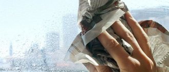
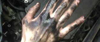
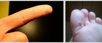
Domestic or industrial injuries often lead to penetration of various bodies into soft tissues (skin, subcutaneous tissue, muscles) - glass and wood fragments, needle fragments and metal particles, plant thorns, etc. Small objects, for example, a wooden splinter located in the superficial layers of the skin, can often be removed independently. However, in order to avoid even greater tissue injury and the development of inflammation and suppuration, such manipulations are best carried out in a medical institution. Removal of foreign objects from soft tissues at the GMS surgery center is performed quickly, painlessly and with minimal trauma.
Example 6. Restoration of teeth 4 and 5, when only one root remains
In the next total work I would like to highlight the 4th and 5th premolars. In general, the girl’s situation was quite complicated initially at the time she contacted me. Both the lower and upper jaws were restored. Let's look at only the top one.
Front 4 teeth
- these are 1.2, 1.1, 2.1 and 2.2 were entirely made of composite material, under which caries developed. And the teeth we were interested in, 1.4, 1.5, 2.4 and 2.5, were covered with metal-ceramic crowns under which there were metal inlays. Today, such structures are rarely used in advanced dentistry, since the same Cerec technology allows you to achieve excellent results with one module “crown + tooth root”, rather than breaking the structure into an inlay and a crown with an additional adhesive layer. In fact, we place a crown at the root of the tooth with the function of the missing root. There will be a separate example on metal tabs a little later.
So, in the photo below, the crowns were removed, the inlays were removed and carious tissue was removed from teeth 4 and 5, leaving healthy roots
:
If only the root remains of a tooth, this does not mean that it cannot be restored. And let this tooth root be pulpless, i.e. dead - such tooth roots feel great in bone tissue and orthopedic structures can be built on them. Using computer technology and 3D scanning, we first restored virtual teeth:
The following photo shows that on one side we place crowns with an inlay function in place of the 4th and 5th teeth, and on the other opposite side we place half-crowns also with an inlay function.
That is, these are single modular designs - veneers with a root part, which are currently the best for the patient
:
Installing veneers with the root part allows you to completely recreate an aesthetically beautiful dentition:
The stage of temporary prosthetics, which allows the patient to see his smile, plays an important role in the very process of its new formation, since the patient understands that the main problems with the restoration of the remaining teeth, and in fact the roots of the teeth, are left “far behind”:
After installing crowns and half-crowns and restoring the front teeth, our patient’s smile was transformed beyond recognition, the so-called. wow effect:
The clinical example described above can be viewed in detail HERE.
Why foreign objects in soft tissue need to be removed
Such a seemingly harmless situation, such as a splinter or a small piece of glass that has penetrated into a leg or arm, can result in a serious inflammatory-purulent reaction, even leading to the development of phlegmon. Systemic infection and intoxication of the body also pose a significant danger. Therefore, in these situations, you need to immediately take adequate measures to remove the foreign object.
But if small splinters and splinters located in the superficial layers of the skin can be removed independently, naturally observing all the rules of asepsis, then you should not experiment on your own with foreign bodies that have penetrated more deeply. In addition, home removal often ends with only the extraction of a fragment of a foreign object, while other particles may go unnoticed. You can avoid such problems by promptly contacting a doctor, who will quickly and professionally remove the foreign body and all its fragments from the tissues.
Examples when only the roots of the teeth in the upper jaw remain
Example 1. Restoration from one root of a front tooth
In this example (presented in full here), the situation was created literally by the patient’s hands. She tried to glue part of the tooth with superglue and, under the influence of dangerous substances contained in the glue, the crown of the tooth became completely unusable.
But the root of the tooth under the crown below remained intact and unharmed.
The length of the healthy part of this tooth root removed all doubts about the question - is it necessary to restore the root? Certainly! - it is possible and necessary. The tooth was restored in 1.5 hours using Cerec technology. And here in the photo is the happy owner of a new restored front tooth:
Example 2. Restoring 3 front teeth, of which only the roots remain
Here is a treatment example where I restored the aesthetics of her smile to a 22-year-old girl. The three front teeth on the upper jaw were completely destroyed. From the upper teeth 1.2, 1.1, 2.1 only
the roots
:
These three teeth were actually an aesthetically unsightly frame made from old fillings. Now we will not analyze the whole case in detail, I would like to draw your attention to the following points:
- the shape of the roots of the teeth and their condition made it possible to restore them
- There was no talk at all about any amputation - removal of the roots of the teeth of the upper jaw and implantation.
Our patient's smile was designed in a computer program:
So, in the photo below you see a general set for restoring our patient’s dentition, and among the modules there are 3 with a crown and a root.
The roots of the upper front teeth were quickly and, most importantly, effectively restored:
and our patient underwent a beautiful transformation and became the owner of a sweet and chic smile:
This is the enormous potential and aesthetic power of healthy tooth roots! Don't rush to delete them)
Example 3. Complete restoration of the smile zone on the front teeth of the upper jaw
This example is actually very popular on the Internet, since the patient is a TV presenter, and as a result of the treatment she found her new smile. Which is extremely important for her profession. I describe the detailed clinical case itself in another article, but here I would like to note the main points related specifically to the restoration of the roots of the front teeth with crowns.
In the picture below we have removed composite restorations and carious lesions. only the roots remain of the two front incisor teeth.
:
Crowns and “crown + root” modules were prepared in the laboratory:
After installation, the smile began to shine with bright colors:
You can see the entire chronology of our patient’s treatment in one large picture, step by step:
Cost of services
The prices indicated in the price list may differ from the actual prices. Please check the current cost by calling +7 495 104 8605 (24 hours a day) or at the GMS Hospital clinic at the address: Moscow, st. Kalanchevskaya, 45.
| Name | Price |
| Removal of a foreign body from the ear, nose, throat | RUB 8,075 |
| Removal of a foreign body with tissue dissection | RUB 11,680 |
| Consultation with surgeon Alexander Givievich Natroshvili | RUB 4,999 |
Dear Clients! Each case is individual and the final cost of your treatment can only be found out after an in-person visit to a GMS Hospital doctor. Prices for the most popular services are indicated with a 30% discount, which is valid when paying in cash or by credit card. You can be served under a VHI policy, pay separately for each visit, sign an agreement for an annual medical program, or make a deposit and receive services at a discount. On weekends and holidays, the clinic reserves the right to charge additional payments according to the current price list. Services are provided on the basis of a concluded contract.
Plastic cards MasterCard, VISA, Maestro, MIR are accepted for payment. Contactless payment with Apple Pay, Google Pay and Android Pay cards is also available.
Western standards of treatment (evidence-based medicine)
Continuous staff development
Regular interaction with leading Russian and foreign medical institutions
Modern medical equipment and advanced diagnostic and treatment methods
Unified standard of service
We work around the clock 24/7/365
Make an appointment We will be happy to answer any questions Coordinator Oksana
Benefits of visiting GMS Hospital
Removal of foreign objects from soft tissues at the GMS Hospital surgical center does not take much time and is performed on an outpatient basis, using local anesthesia and modern surgical equipment. By contacting us, each patient receives:
- fast, qualified surgical care without queues or delays;
- treatment of wounds and injuries of various types;
- non-hazardous and painless removal of a foreign object of any nature;
- in the presence of a serious traumatic injury, hospitalization is possible;
- the surgery center is equipped with the necessary equipment for rapid diagnosis and surgical treatment of foreign bodies in soft tissues;
- The operation is performed within an hour after treatment on an outpatient basis.
The center’s specialists have extensive experience in performing such interventions and quickly remove an object of any nature, using modern equipment and safe anesthesia. To make an appointment, please call the reception desk or leave a request on the website.
Indications for surgery
You need professional help when inserting any object into soft tissue due to injury. Do not delay your visit to the surgeon if the following situations occur:
- inability to independently remove a foreign object;
- development of bleeding due to damage to the vessel by a foreign body;
- when palpating the location of a foreign object, severe pain is felt;
- infiltration, inflammation or suppuration has occurred in the area where the object was introduced;
- the wound does not heal, a purulent fistula has formed;
- loss of sensitivity is noted at the location of the foreign object.
There is no need to endure pain and delay treatment. All this can end in wound suppuration and general blood poisoning. To avoid complications and quickly forget about the incident, promptly contact a surgeon who will quickly and painlessly carry out all the necessary manipulations.
Symptoms of eye damage
The following types of symptoms indicate that scale has gotten into the eye:
- sudden onset of pain;
- redness of the protein membrane;
- increased photosensitivity;
- spontaneous closure of the eyelids;
- blurry vision;
- burning in the eyes;
- tearfulness;
- swelling of the eyelids.
These are the main signs of damage. Only an ophthalmologist can make an accurate diagnosis.
Preparation, diagnostics
To safely and accurately remove foreign objects from soft tissue, you need to determine its location as accurately as possible. For this purpose, the following techniques are used:
- radiography (in the presence of radiopaque bodies, for example, metal);
- transillumination – in the presence of glass, plastic objects, wood, etc.;
- Ultrasound of soft tissues.
If the intervention involves tissue incision, preoperative preparation includes a set of blood tests (biochemistry, general, sugar, coagulability, group and Rh factor, hospital infections). In situations where part of the foreign object is located above the surface of the skin and can be easily removed without resorting to incisions, no preparation is required.
Examples of canine restoration when only the root remains of the tooth
Example 4. The pin in the root of the lower tooth was removed, and the canine itself was restored
In this clinical case, I was restoring the lower canine, and the task was to save the root of the tooth and restore the coronal part of the canine, which was almost destroyed by caries. The anterior wall of the lower canine was made in the form of a filling, and it fell apart from the cutting edge.
As you can see in the photo, an anchor pin was installed inside the tooth - this is a pin that is screwed into the canal to strengthen the tooth. The tooth under the filling and the root of the lower canine were affected by caries. When the carious tissue was removed, only the root and a small part of the wall
:
You and I remember the main topic of our article, which tells us the following - if the tooth root remains, it can be completely restored. And this is not the first time that the unique Cerec technology has come to our aid. Using it and digital scanning, we restored the missing tooth module, which also included the root part:
Next, we restore the lower front tooth using a CEREC inlay and get an excellent result - the lower canine is completely restored:
Example 5. The root of the tooth under the crown of the upper canine has not rotted, and therefore the tooth is 100% restored
In this case, there was a total restoration of the dentition (veneers and crowns) with the replacement of worn-out crowns on the front teeth. One of the upper canines under the crown was seriously affected by caries, but its root was practically healthy - only a small part of the tooth root was damaged, see the following photo:
The missing “crown + root” module and new crowns were manufactured in a laboratory. In general, everything is in a smile, it looks just great:
And the owner of a new beautiful smile herself cannot hide her admiration:
All the main tasks in this total work were successfully solved, but the main thing you should pay attention to is that we managed to save the tooth root here too.
How is the operation performed?
In most cases, surgery is performed under local anesthesia and takes a few minutes. General anesthesia is used only for large operations. The doctor uses a scalpel to expand the wound canal to the required depth, grasp and remove the foreign object. After this, the wound cavity is treated with an antiseptic, sutures are applied, drained (if necessary) and everything is covered with a sterile bandage. In almost all cases, the patient is administered antitetanus serum and prescribed a short course of antibiotic therapy to avoid the development of complications.
You have questions? We will be happy to answer any questions Coordinator Tatyana
Example 9. Restoring the bottom six “from the root”
In this case, I would like to simply dwell on the moment of restoration itself. The patient is older, over 50 years old. And he felt the need to restore the chewing group of teeth. The picture shows the lower 6th tooth, of which practically only one root remains:
The tooth was restored in 1.5 hours
using Cerec technology. The very speed of tooth restoration in one visit to the doctor and the patient’s ability to immediately, as they say, use it in chewing gives a 100% head start on any existing method of tooth restoration. It works really well - you can see for yourself.
Example 10. Restoring the bottom seven
And I end my examples with the classic option of restoring the seventh lower tooth, of which only the root part remains. Removal of the lower 7th root of the tooth was not required; Cerec technology again proved to be excellent:
After modeling in 3D, manufacturing the module itself only took about 20 minutes, after which it was successfully installed on the remaining root:
Features of the rehabilitation period
In order for healing to proceed quickly and without problems, after the intervention you must follow medical recommendations:
- regularly treat the surgical wound and change dressings;
- minimize the load on the injured area;
- exclude thermal procedures (hot bath, saunas, steam baths);
- Do not visit the pool or bodies of water.
To make an appointment with GMS Hospital specialists and remove a foreign object, please call or fill out the feedback form on the website.